
- Home
- Dipartimento
- Ricerca
- Didattica
- Post Lauream
- Trasferimento della conoscenza
- Come fare per
Alumni Colloquium @ DF - S. Olivo - Seeing invisible things with x-rays
Tipologia evento:
home
Sede:
Trieste
You are all familiar with x-ray procedures, e.g. what they do to you if you go to the hospital because you have fallen off your bike and think you might have broken your wrist. Actually the use of x-rays in science and society is much more widespread: as well as in medicine, x-rays are used in industrial testing, materials science, biology, security inspections, cultural heritage preservation – the complete list would probably take up the entire length of this page.
That said, technological developments on sources and detectors aside, x-ray imaging has been performed in pretty much the same way since Roentgen’s discovery of x-rays 120 years ago: by exploiting differences in x-ray attenuation. This is often sufficiently reliable – however when attenuation differences are small, either because you are trying to distinguish materials with similar attenuation coefficients (e.g. tumours in soft tissue), or simply because the features you are targeting are thin, then this conventional approach breaks down, and you are left with practically no image contrast.
For this reason, X-Ray Phase Contrast Imaging (XPCI) has been the subject of intensive research over the last ~20 years, and it is widely believed that its use can transform all applications of X-ray imaging. In XPCI, contrast arises from refraction/interference effects instead of absorption, which leads to the visualization of features classically considered “x-ray invisible” and, more generally, to a significantly enhanced visibility of all details in an image.
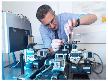
While until recently it was believed that XPCI was restricted to synchrotron environments, the UCL group has developed a method that works with conventional x-ray sources, hence opening the way to its translation into mainstream use, as well as making XPCI available to a potentially much wider user base.
More recently, microscopic and CT implementations of the lab-based method were also developed, as well as a “dark-field” phase-based approach which provides additional, complementary information on the sample at lengthscales below the resolution of the imaging system.
Moreover, when implemented with synchrotron sources, the UCL methods outperforms others in terms of phase sensitivity, a feature which is currently being investigated in more detail as it can potentially enable scientific applications previously considered inaccessible.
In this talk I will introduce the basic principles of XPCI, explain how the UCL method works, and discuss a number of possible applications – both in terms of translation to “real-world use” and enabling access to new scientific areas.
Luogo:
Lecture Room A, via Valerio 2
Ultimo aggiornamento: 20-11-2015 - 17:50