Advanced X-ray imaging research from UniTS featured in Physics World
Research in the field of advanced X-ray imaging, led by researchers from the University of Trieste, has been the subject of a recent interview on Physics World, the magazine of the Institute of Physics, one of the largest physics associations in the world.
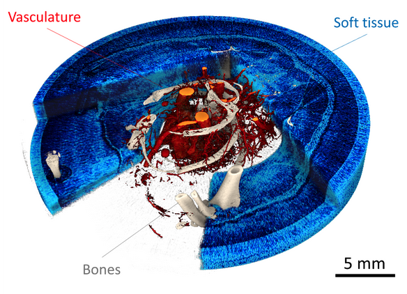
The research, published in Physics in Medicine and Biology (Brombal et al. 2024), concerns the first-of-its-kind combination of two cutting-edge imaging methods: spectral tomography and phase contrast imaging. The former allows for quantifying material density in three dimensions, while the latter allows for increased visibility of soft tissues, which are poorly visible with conventional techniques. The simultaneous use of the two techniques allowed for acquiring high-resolution (0.02 mm) and high-concentration sensitivity (few mg/ml) quantitative three-dimensional maps.
The study, which involved an international team of researchers from University College London (UK) and the Helmholtz Zentrum in Munich (Germany), as well as staff from the Departments of Physics (DF) and Engineering and Architecture (DIA) of UniTS, was conducted at the Elettra Synchrotron in the beamline dedicated to medical physics applications SYRMEP and co-funded by the National Institute of Nuclear Physics (INFN).
As reported in the interview, the next step in the research, supported by PNRR funds, will be translating this technique into a compact laboratory and its application to scientific cases of biomedical interest, first and foremost research on the development of osteoarticular diseases.
In the picture: Image taken from Brombal et al. (2024) showing a high-resolution 3D rendering clearly displaying the vascular, bone, and soft tissue components of the sample.